Projekty »
Mechanisms responsible for brain defence and protection
| :: Projekt UP261 (Szczegóły) | |
| Terminy |
| Czas trwania projektu: 2 godz. (90 min.) |
Miejsce realizacji: Collegium Veterinarium (236)
Adres: Lublin, ul. Akademicka 12
The brain is the most important part of the central nervous system and at the same time the paramount organ in humans and animals. It ensures the condition of the whole organism by controlling the activity of all organs and maintaining the proper homeostasis. It is also responsible for higher nervous functions such as cognition, memory and learning. The brain may be exposed to different harmful physical, chemical or biological factors. Due to this fact, the brain was secured by different protective and defensive mechanisms. The skull and the meninges protect the brain against mechanical factors. It is protected against chemical factors by a tight blood-brain barrier. Whereas the biological factors are fought off by glial cells: astrocytes and microglia. Glia can modulate the functioning of the immune system, which also affects the immunity of the entire body. In case of damaging the brain, glial scars may be formed or, according to recent studies, new nerve cells may appear. Neurogenesis plays an important role in filling defects, e.g. in the hippocampus, and therefore it can restore learning and memory abilities after an injury. The aim of the presentation is to present various protective and defensive mechanisms against dangerous factors within the brain.
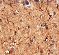
Each participant of 20th Lublin Science Festival will personally watch histological slides under the light microscope and analyse the structures of central nervous system. The microscopic analyses will be preceded by an introduction with use of multimedia presentation.

